

Contributors
Supported by the National Institute of Neurological Disorders and Stroke, National Institutes of Health, under grant No. NIH-Grant R37 NS065434. BRIDGE-UP Program funding is provided by the National Institute of Allergy and Infectious Diseases, under grant No. R25AI170381
Preview
Description
Poster presented at the Annual Biomedical Research Conference for Minoritized Scientists (ABRCMS) in Pittsburgh, Pennsylvania
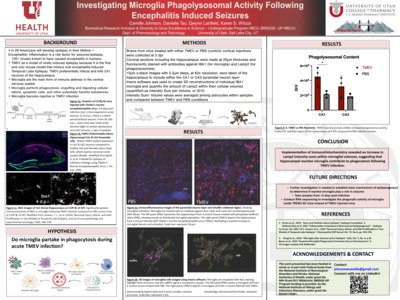
This poster investigates the role of microglia in phagocytosis during Theiler’s murine encephalomyelitis virus (TMEV)-induced epilepsy, a viral model for temporal lobe epilepsy (TLE). TMEV preferentially infects hippocampal CA1 neurons, leading to acute and recurrent seizures. Microglia, the central nervous system’s primary immune cells, become reactive during TMEV infection. This study examines microglial activation and phagocytic activity using immunohistochemistry and 3D imaging to assess the microglial response in the hippocampus. Results show increased microglial density and phagolysomal activity in TMEV-infected mice compared to controls, suggesting microglia contribute to tissue repair during the inflammatory response. Future research will explore the role of reactive microglia in epileptogenesis, with plans for RNA sequencing and further investigations using TREM2 knockout models.
Format
PNG
Keywords
Microglia, Phagocytosis, Theiler's Murine Encephalomyelitis Virus (TMEV), Temporal Lobe Epilepsy (TLE), Hippocampus, Reactive microglia, Epileptogenesis, Seizures, Immunohistochemistry, 3D Imaging, Phagolysomal activity, Microglial activation, Inflammation, TREM2 knockout models, RNA sequencing, Mouse model, CA1 neurons, Central nervous system immune response, Tissue repair, Neuroinflammation